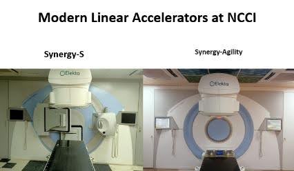
SYNERGY-S® & SYNERGY-AGILITY®
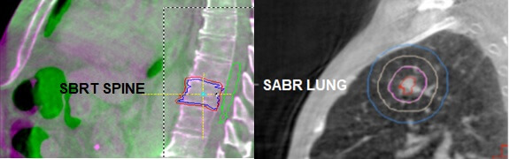
Stereotactic Body Radiotherapy (SBRT) and Stereotactic Ablative Radiotherapy (SABR) Techniques
by Elekta is an image guide robotic linear accelerator that combines high conformance beam shaping with exclusive 3D adaptive image guided radiation therapy (IGRT) / Volumetric Modulated Arc Therapy (VMAT). It is dedicated for intra-cranial and extra-cranial applications.
Decades have contributed to improved results and wider applications of radiosurgery. The role of radiosurgery has expended beyond its initial applications. Synergy-S is the ideal extra-cranial complement to Leksell Gamma Knife as combination of both creates stereotactic center of excellence.
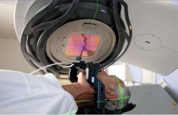
It works like Gamma Knife having a head frame, body fix imaging, image planning and special modality of self-imaging planar fluoroscopic event/ and 300-ray volume imaging. Its own table to confirm the target and then treatment either radiosurgery or radiotherapy over confirmed target. It has maximum accuracy in advanced stereotactic radiosurgery with reference to spinal applications like spinal metastasis. Its fluoroscopic image modality prior to treatment helps to minimize the risk of set-up error by identifying critical structures during treatment, thus compensating organ motion like heart, lungs, larynx and abdominal structures.
Having Synergy-S multi-leaves (micro-macro) collimator system in conjunction with powerful software driven 2D & 3D image-guided accuracy, enables oncologists to apply SRS/SBRT effectively for small and large field lesions.
Indications:
- Intracranial Indications:
- Large tumors, tumors closely lying near to critical organs, tumors embedded into the organ at risk or microscopic disease, etc.
- Benign:
- Meningioma, Pituitary Adenoma, Acoustic Schwannoma, AVM, Cavernoma, Functional disorders, Psychiatric disorders, etc.
- Malignant:
- Brain Metastases, Recurrent Gliomas, etc.
- Extra Cranial Indications:
- SBRT is now a standard procedure for many of the extra-cranial regions including: Spine, Lung, Liver, Prostate, and Oligometases (New concept with cure or radical treatment with SBRT/SRS in stage –IV Cancers.
Volumetric Modulated Arc Therapy (VMAT)
It was introduced in 2007 and was described as a novel radiation technique that has continuous modulation of MLCs (field shaping), along with the dose rate and gantry speed rotation utilized to deliver highly conformal dose distributions simultaneously, in a minimal time period with reasonable MUs to be delivered.
VMAT carries an advantage over IMAT in terms of greater degree of freedom that increase the capability of beam intensity modulation. Teoh et al. published comprehensive review article on VMAT in 2011.
VMAT is becoming an increasingly utilized radiation technique. It has proved as a novel and emerging technology with freedom of selection of number of arcs and some other features making it more efficient and faster in terms of treatment time and MUs delivery, its ability to spread low dose to a wide area of normal tissue, ability to deliver complex treatments with coplanar or non-coplanar single or multiple arcs make it a unique technology. The risk of secondary malignancy in VMAT should be lower as VMAT generally uses fewer monitor units (MU) compared with conventional fixed field IMRT. Macchia et al, published a detailed review of VMAT and its clinical use for various body sites in 2017, this study described that VMAT has been used mostly for brain tumors, head & neck, thoracic cancers, GU cancers, GI cancers and SBRT for oligometastases.
VMAT-SIB is a popular technique that allows treatment of several volumes with different dose prescriptions called simultaneous integrated boost (SIB) is conveniently executed with VMAT technology that results into delivery of high biological effective doses to the target and reduction of the dose to the surrounding normal tissues and improvement of the toxicity. Macchia et al, concluded that the clinical use of VMAT is less documented, but VMAT-SIB and VMAT-SBRT is an effective and safe technique for various cancers of the body. More clinical data will emerge by the time as the numbers of patients are increasing across the world.
VMAT-SRS is a reliable therapeutic modality of SRS based upon the existing diametric research on its safety and benefits particularly in multiple brain metastases. They considered VMAT similar to the “non-VMAT” approach in terms of treatment plan acceptability (conformity and heterogeneity), treating multiple lesions and offering frameless radiosurgery treatments under image guidance.
In 2008, Gamma knife & Linac based Stereotactic Radiosurgery/Radiotherapy was established at Neurospinal & Cancer Care Institute (NCCI) Karachi by Prof. A. Sattar M Hashim.
Our group emerged as a pioneer of stereotactic radiosurgery (SRS) /stereotactic body radiotherapy (SBRT) in Pakistan. Initially we were using various forms of IMRT: fixed beam, step and shoot, forward and inverse IMRT from 2008 till 2013.
Later we were able to acquire a license to use VMAT technology in Pakistan and since 2014 we are using VMAT, VMAT-SIB and VMAT-SRS/SBRT techniques on a regular basis.
SYNERGY-S / SYNERGY-AGILITY
Along with Synergy-S, Another New Linear Accelerator “SYNERGY-AGILITY has already been Installed at NCCI to carry out Stereotactic Body Radiotherapy.
Revolutionary Accuracy In Treatment Delivery
Synergy-S is the only fully digitally controlled linear accelerator available for extra-craniaI stereotactic applications. It is the first system of its kind with 3D imaging of the target area at the point of treatment delivery. We call this 4D Adaptive IGRT – high-resolution imaging in three dimensions of physical space and the tooth dimension of time.
Dose Control And High Conformance
Synergy-S delivers high dose rates with exceptionally fine control and precision. The system enhances avoidance of critical structures during beam delivery with its innovative 3D imaging technology. This technology allows beam adaptation for individual treatment sessions to achieve high precision and excellent target conformance, plus, digital controls which maximize the accuracy of dose delivery without compromise.
Synergy- S offers with stable dose/emu calibration and the most accurate IMRT/VMAT delivery available.
Multi-Modality Energy Flexibility
Synergy-S offers an unprecedented selection of therapeutically effective X-ray beams. With the flexibility to select a higher energy X-ray beam than AMA, physicians can optimize the dose distribution by choosing energy appropriate to the site and depth of the lesion.
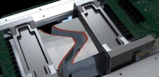
Intelligent Beam Shaping
Agility is the next generation, high resolution beam shaping solution from Elekta. Built on a strong understanding of the factors that are critical to patient plan optimization and treatment delivery, Agility is designed to meet the needs of modern radiotherapy facilities. Agility offers excellent clinical flexibility and efficiency, while ensuring that patient safety and comfort remain a priority.
Rapid leaf
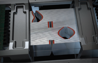
40 x 40 cm field size.One hundred and sixty Inter digitating leaves with 5 mm width at isocenter. Integrated digital control of leaves and dynamic leaf guides. Accurate leaf positioning with Rubicon optical technology. Rapid leaf speed. Extremely low leaf transmission (<0.5%). 45 cm isocentric clearance.
Shorter Treatment Times
The high leaf and diaphragm speed provided by Agility increases the speed of delivery and allows higher dose rates to be used for more effective modulation. Not only does this help to improve plan quality, but it also shortens treatment times considerably. Shorter treatment times mean that more patients can be treated in a working day.
Multifunctional
Agility is ideal for the treatment of large or small field shapes and for the delivery of static or dynamic treatment plans for conventional or stereotactic techniques. You can even deliver off-center, non-coplanar treatment plans.
Agility offers high resolution beam shaping, including interdigitation, across the 40 x 40 cm field size. Synchronized intelligent dynamic leaf guide movement and individual leaf movement achieves enhanced leaf speed and removes the need for a split field for seamless delivery of each prescribed beam shape.
These features combined with a market leading 45 cm iso-centric clearance ensure that your Linac is truly multi-functional.
Rubicon Optical Positioning System
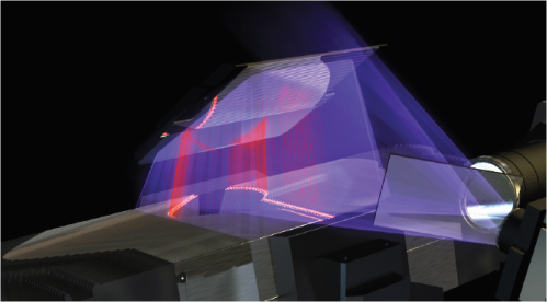
The robust and reliable “Rubicon optical positioning system” provides valuable real-time assurance of accurate leaf positioning. Agility’s Rubicon optical technology offers advanced real-time leaf monitoring and positioning. Ultraviolet light from an LED source produces infrared fluorescence when it falls on the ruby tips of the multileaf collimator leaves. This infrared fluorescence, detected by an infrared camera, is used to reliably monitor and accurately position the leaves and can be viewed in real-time on the linear accelerators display screen.
Inherent Safety Supervisor
In addition, Integrity™, the fully integrated Elekta digital control system, provides: Continuous monitoring and control throughout treatment for confidence in beam shaping and treatment delivery. Verification that the field being delivered matches the prescribed treatment parameters. Inherent safety features that supervise and check that all systems are operating correctly.
Lowest Leaf Transmission
The transmission and penumbra measurements indicate a significant improvement on the published data for comparable equipment. In particular, leaf transmission is extremely low, less than 0.5%. Cosgrove et al (2009). Agility has exceptionally low leaf transmission at less than 0.5% across the entire field1. This is important for a number of reasons:
It reduces unwanted dose to organs at risk and healthy tissue. Low integral dose reduces the risk of inducing secondary tumors. It reduces off axis dose when performing off-axis beam shaping. It improves treatment plan quality.
High-Resolution Beam-Shaping
Synergy S and Synergy-Agility includes an integrated multi leaf collimator with a generous 40cm x 40 cm field size. The field comprises 160 individually controlled leaves. Physicists can create a range of finely shaped, high-resolution fields within one field simultaneously. This contributes to patient safety and improved conformal avoidance of critical structures.
Beam Angle Flexibility
Synergy S and Synergy-Agility features a small diameter 62cm treatment head which in combination with the best isocenter clearance, maximizes clearance around the patient and contributes to the wide variety of treatment approaches possible, including non-coplanar treatments. This enables Synergy S and Synergy-Agility to deliver radiation from angles impossible to replicate with other systems. Synergy S and Synergy-Agility can also radiate from underneath the couch for spinal applications, with the patient in the supine position.
Complete Quality Assurance
Integrated quality assurance (QA) tools ‘whole a kV/MV phantom to maintain excellent image quality and automated multi-leaf calibration software for the beam modulator that utilizes iView-GT MV images. This turnkey QA package enables the physics team to maximize efficiency and maintain the highest levels of device precision.
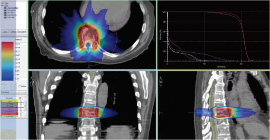
ABSOLUTE ACCURACY IN PLANNING- MONACO 5.11 – TPS
Synergy S and Synergy-Agility integrates a suite of sophisticated software tools to provide unprecedented target accuracy and help improve patient outcomes. Monaco-5.11- Treatment Planning System (TPS) reduces complexity and enables Physicists to create high-precision plans that are easy to deliver and verify. It supports all linear accelerator clinical techniques.
Stereotactic Body Frame
Stereotactic Body Frame enables clinicians to localize turners precisely in a 3D stereotactic coordinate system, increasing the accuracy of radiation delivery. It allows high dose treatments with minimal exposure to surrounding healthy tissues, establishing a reliable basis for hypo fraction. With the Stereotactic Body Frame, neurosurgeons can reproducibly localize isocentric target coordinates during planning and treatment. The system includes built-in reference indicators (fuducial markers) for CT and MR determination of target coordinates.
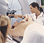
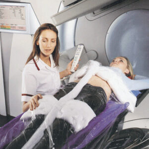
Active Breathing Coordinator
Active Breathing Coordinator allows clinicians to pause a patient’s breathing at a precisely indicated tidal volume and coordinate radiation delivery within this pause. This simple, portable system helps achieve repeatable breath hold for accurate delivery. In addition Active Breathing Coordinator reduces overall treatment time compared to alternate gated systems. It can be used with CT-simulation to enable reproducibility and increase precision of treatment. Active Breathing Coordinator offers a simple solution for controlling internal motion without the need for external markers.
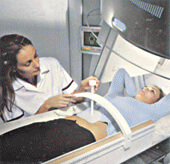
Body FIX RT
Body fix RT immobilizes firmly and gently throughout imaging, simulation and treatment. It is ideal for use with Synergy-S, In conjunction with Hexa POD and minimizes involuntary patient movement during imaging and therapy. BodyF1X RT is composed entirely of radio-translucent materials compatible with CT, MR, PET, SPECT and ultrasound modalities.